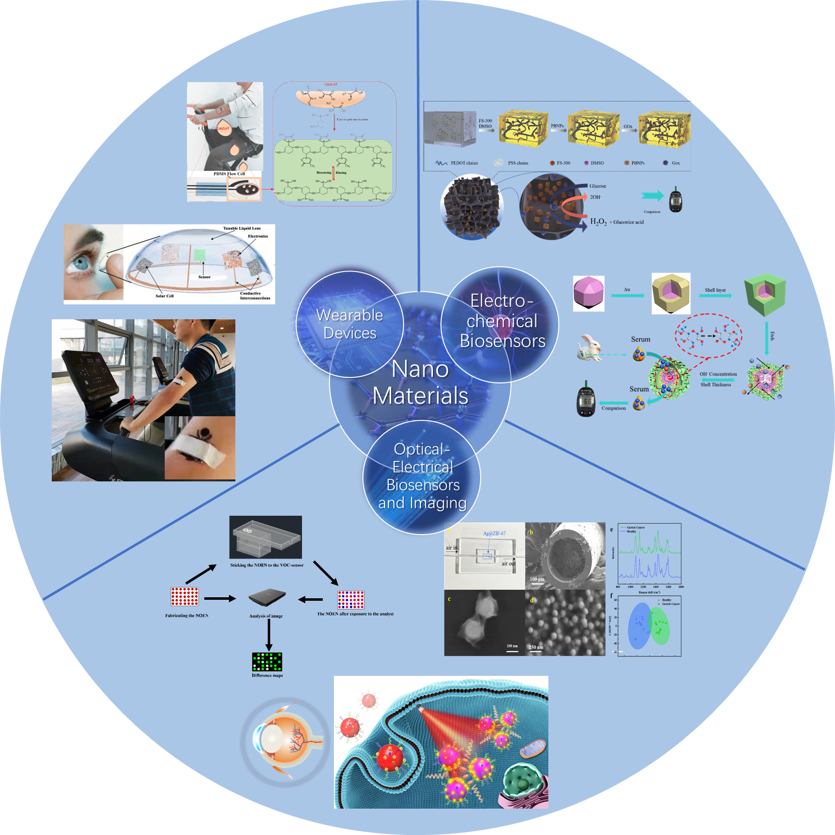
The main research directions of this research group are as follows:
1. Biomimetic biosensor based on nanomaterials;
2. Bio-imaging with nanoprobes based on SPR and SERS;
3. Development of electrochemical biosensors and spectra imaging systems;
4. Development of wearable sensors;
5. AI for bio-imaging.
1. Preparation of nanomaterials and their application in (wearable) electrochemical sensors
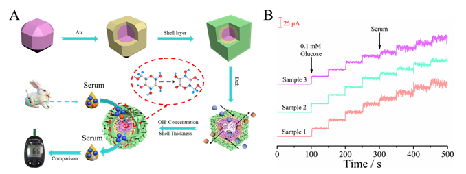
(1) The as-prepared sandwich and core-shell structure of Cu(OH)2@Au@Co(OH)2 presented an ultrahigh sensitivity to glucose. The sensitivity is as high as 3427μA mM-1cm-2. Using rabbit serum as the real sample, the test results are almost the same as that of commercial blood glucose sensor, with good accuracy.
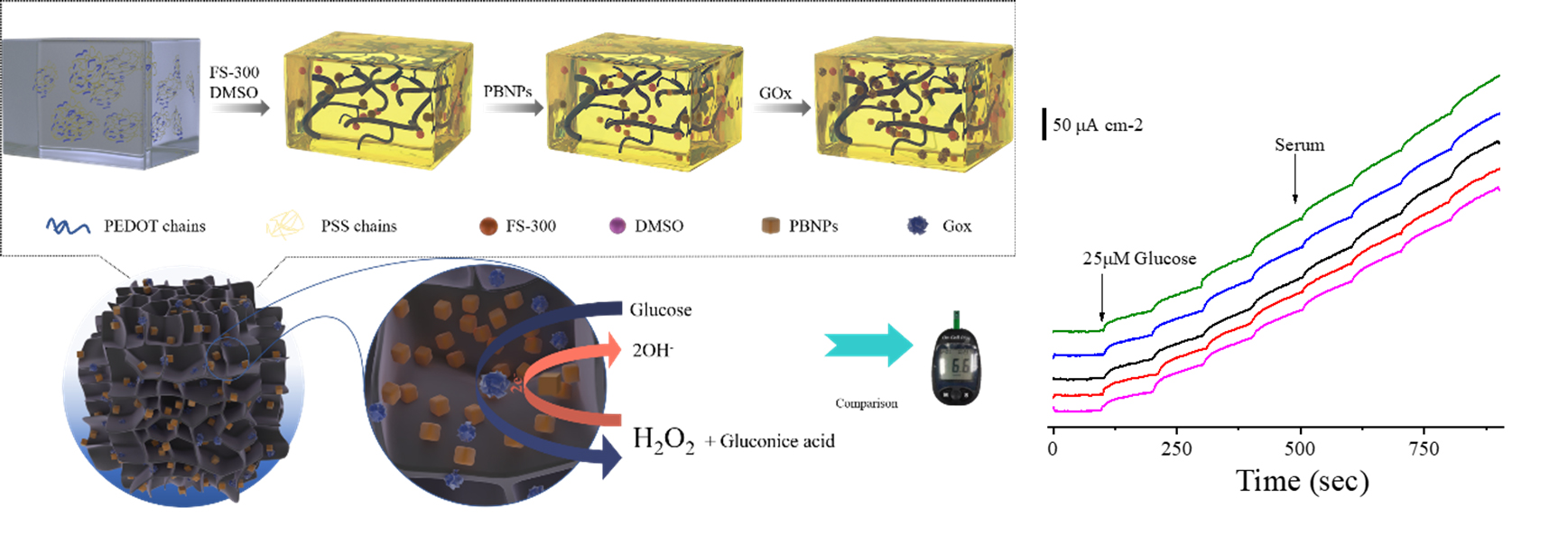
(2)The prepared conductive hydrogel composite sensing material has excellent performance for glucose detection in serum.
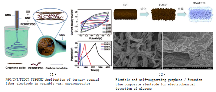
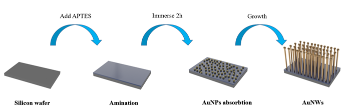
(3) The preparation and photoelectric sensing applications of metal organic framework materials, two-dimensional materials and nano superlattices.
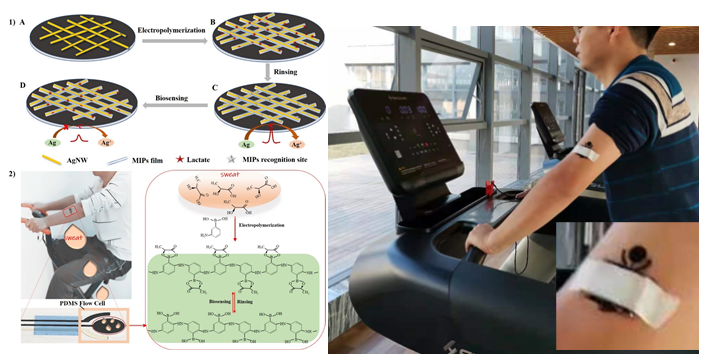
(4) Wearable sweat lactic acid sensor, based on silver nanowires and other flexible electrode materials electrode materials for high sensitivity on-body detection of sweat lactic acid and other molecules.
2. Cancer diagnosis and screening based on Raman technology, microbial detection
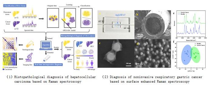
(1) Based on Raman spectroscopy technology and artificial intelligence algorithm, the rapid, convenient and high sensitivity diagnosis and identification of liver cancer tissues and adjacent tissues as well as different types of liver cancer tissues are realized. The boundary imaging of liver cancer and tissue section imaging at subcellular level are realized by using Raman spectroscopy imaging technology.
(2) Metabolic differences caused by cancer can cause differences in volatile organic compounds (VOCs) in human exhaled air. We first used GC-MS to determine the exhaled gas markers of gastric cancer patients and healthy people, and then used the prepared Ag@ZIF-67-based materials for VOCs collection and measurement. The flow-through surface-enhanced Raman sensor successfully distinguished gastric cancer patients from healthy people.
(2) Metabolic differences caused by cancer can cause differences in volatile organic compounds (VOCs) in human exhaled air. We first used GC-MS to determine the exhaled gas markers of gastric cancer patients and healthy people, and then used the prepared Ag@ZIF-67-based materials for VOCs collection and measurement. The flow-through surface-enhanced Raman sensor successfully distinguished gastric cancer patients from healthy people.
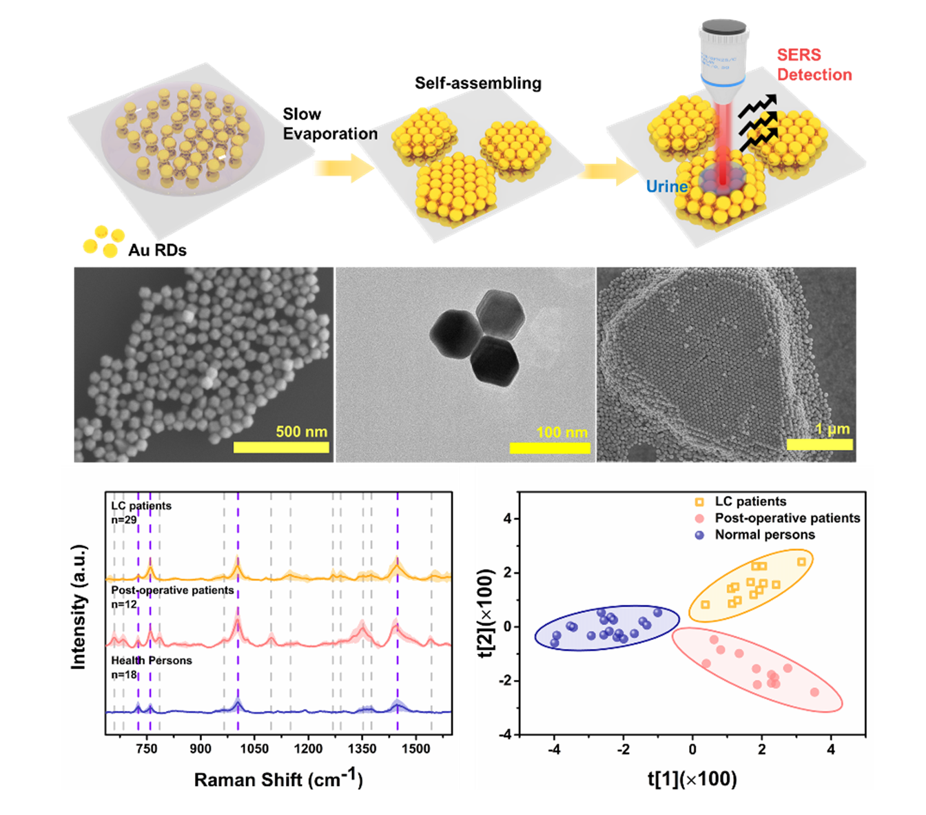
(3) Using the Raman enhanced substrate prepared by nano superlattice, we can distinguish the urine of patients with lung cancer before and after operation and normal people, and assist in lung cancer screening and postoperative recovery detection.
(4) A SERS signal enhancement substrate was prepared to capture and identify ophthalmic pathogenic microorganisms.
3. Research on the detection and imaging of neurotransmitters in nerve cells and tissues based on SERS
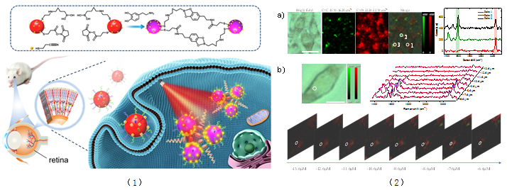
(2) a) Raman imaging and SERS spectra of multiple PC12 cells. The light field pattern of multiple cells, the Raman images of 1570-1620cm-1 and 2220-2270cm-1 channels and their combination, and the Raman spectra of points 1, 2 and 3. Scale: 20μm. b) Three dimensional imaging of a single PC12 cell. The bright field pattern of a single PC12 cell and the SERS spectra of different layers in the white circle, as well as the Raman imaging of 2220-2270cm-1 channels in different layers in the Z direction of the same cell. Scale: 10μm.
4. Colorimetric sensor for cancer diagnosis
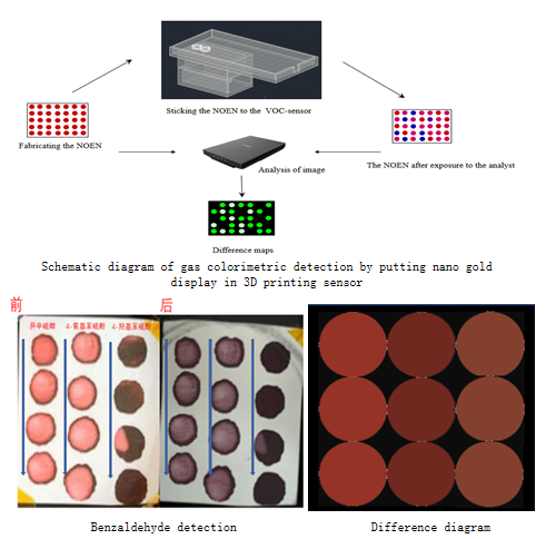
Using nanomaterial array, the sensor of exhaled gas VOC detection was prepared, for noninvasive lung cancer and gastric cancer screening.
5. Biosensors on contact lens
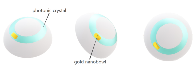
Contact lens are used as the biosensor matrix for the diagnosis of ophthalmic diseases. When the photonic crystal is combined with contact lenses, the increase of intraocular pressure leads to the change of lattice spacing of photonic crystal, which makes the Bragg diffraction peak shift, and the color change is recorded by fiber spectrometer and smartphone, so as to achieve the real-time monitoring of intraocular pressure. In combination the contact lens with the gold nanobowl substrate, we are able to measure the biomarkers for ophthalmic disease based on SERS.
6. Smartphone SPR imaging system, and SPR microscopy
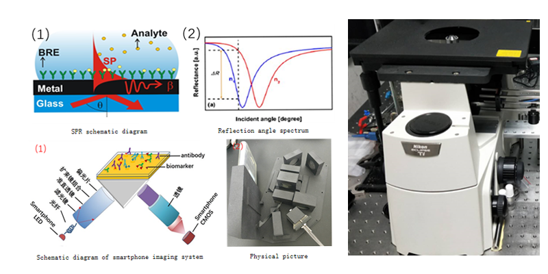
Smartphone SPR imaging system was developed for molecular detection and analysis of molecular interactions. In addition, the labeling free imaging of single nanoparticles and molecules interactions are achieved based on SPR microscopy.
------------------------------------------------------------------------
Current Grants
主持的项目:
【1】国家重点研发计划项目(课题),2017YFC0111300,
【2】国家自然科学基金面上,22274116
【3】国家自然科学基金青年基金,21605116
【4】浙江省杰出青年科学基金,R19H180001
【5】浙江省公益技术应用研究计划项目,2017C33193,
【6】浙江省千人配套经费,ZJQRWZ20191001,
【7】温州生物材料与工程研究所重大科研启动项目,WIBEZD2014004-02,
【8】温州医科大学科研启动项目,89219031
【9】温州市领军型人才创新创业项目,RX2016005,
【10】温州市公益性科技计划项目,Y20160065,
【11】温州市龙湾区科技发展计划项目,2016YG15。
【12】中国科学院大学温州研究院应急项目,WIUCASYJ2020002